The Throat Mass
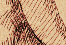
There are a number of small muscles in the area of the throat between the sternocleidomastoids. These muscles are related to speech, swallowing, and breathing. The throat mass has two distinct planes: a horizontal plane under the mandible, and a vertical plane on the front part of the neck.
These planes intersect at the hyoid bone, which is the crowning form of the windpipe. On the anterior surface of the windpipe there are two major forms: the thyroid cartilage and the thyroid gland. The thyroid cartilage is the prominent Adam's apple in the male. On the female the cartilage is flatter and the gland is fuller, making them more evenly-sized on the surface. On the male, it may be possible to see an additional form at the top of the gland called the cricoid (CRY-coid
) cartilage; this is usually covered by the gland in the female.
Most of the plane below the jaw is formed by mylohyoid (MY-loh-high-oid
), which extends from a long line on the mandible to the hyoid bone, with both sides meeting at a tendinous midline.
The anterior bellies of digastric (die-GAS-tric
) run from the hyoid bone over the surface of mylohyoid to attach on the mandible, at roughly the point of the corners of the chin. They may be visible as a pair of columnar bands.
On the vertical plane of the throat, the major form is sternohyoid (STERN-oh-high-oid
). Sternohyoid runs alongside the windpipe from the clavicle to the hyoid bone, nearing the midline as it does so. If visible, it will be seen as a flat band on either side of the windpipe, forming the inner walls of the pit of the throat.
These muscles can usually only be seen when the neck is strongly tensed, and the artist is hardly ever called upon to draw them. It is nevertheless helpful to know their structure for rendering the forms of the throat, whose complexity can easily become a muddle on paper.
