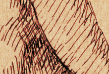
Tibialis Anterior
| Pronuncation | tib-ee-AL-iss an-TEER-ee-or |
| Derivation | On the tibia. |
| Origin | Upper lateral and anterior surface of tibia. |
| Insertion | Bottom surface of the first metatarsal and the cuneiform bone of the foot. |
| Action | Bends the joint of the ankle, drawing the top of the foot upward; turns the bottom of the foot inward. |
Most of tibialis anterior lies on the upper half of the tibia, and extends slightly in front of the bone. Below this a tendon can be easily seen descending diagonally across the tibia and down the medial side of the foot.
Its prominence in front of the tibia makes its forward edge visible in a medial view of the lower leg. In this view, one can see the tibia as the largest continuous area of subcutaneous bone on the body. (This area can be felt as a hard, wide line from the knee to the ankle on this side.)
Immediately adjacent to tibialis anterior is extensor digitorum longus. This muscle repeats the lateral curve of tibialis as a slender band. At the ankle, its tendon splits into the four tendons that extend over the instep to toes #2-5, which this muscle raises.
This slender band is given some apparent extra length by peroneus tertius, which lies below on the fibula. A short span of its tendon may be visible to the outside of the little-toe tendon of extensor digitorum longus. This muscle turns the bottom of the foot laterally.
